COVID-19–associated Acute Hemorrhagic Necrotizing Encephalopathy: CT and MRI Features
RADIOLOGY
COVID-19–associated Acute Hemorrhagic Necrotizing Encephalopathy: CT and MRI Features
Neo Poyiadji, Gassan Shahin, Daniel Noujaim, Michael Stone, Suresh Patel, Brent Griffith
Published Online:Mar 31 2020
https://doi.org/10.1148/radiol.2020201187
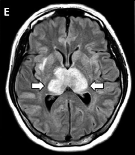
Figure 2d: MRI images demonstrate T2 FLAIR hyperintensity within the bilateral medial temporal lobes and thalami (A, B, E, F) with evidence of hemorrhage indicated by hypointense signal intensity on susceptibility-weighted images (C, G) and rim enhancement on postcontrast images (D, H).
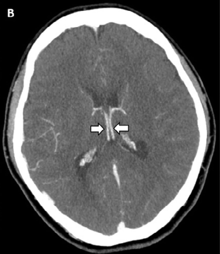
Figure 1b:A, Image from noncontrast head CT demonstrates symmetric hypoattenuation within the bilateral medial thalami (arrows). B, Axial CT venogram demonstrates patency of the cerebral venous vasculature, including the internal cerebral veins (arrows). C, Coronal reformat of aCT angiogram demonstrates normal appearance of the basilar artery and proximal posterior cerebral arteries.