


Hypoxaemia related to COVID-19: vascular and perfusion abnormalities on dual-energy CT
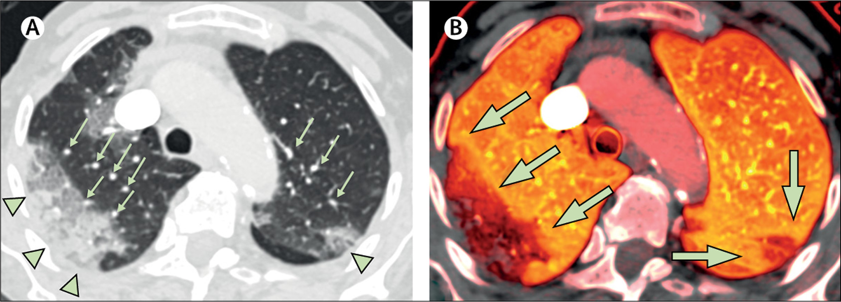
Patient 1, an 87-year-old woman with a history of fever and cough for 5 days, was found on the floor of her nursing home. On admission to hospital, the patient required a non-rebreather mask with a flow rate of 15 L/min to maintain an oxygen saturation of 85%; intubation was not pursued as the patient’s status was comfort measures only. (A) There is a large area of peripheral ground-glass opacity and consolidation within the right upper lobe and smaller ground-glass opacity in the posterior left upper lobe (green arrowheads), which are accompanied by dilated subsegmental vessels proximal to, and within, the opacities (green arrows). (B) The accompanying image of pulmonary blood volume shows corresponding wedge-shaped areas of decreased perfusion within the upper lobes, with a peripheral halo of higher perfusion (green arrows). COVID-19=coronavirus disease 2019.
https://www.thelancet.com/journals/laninf/article/PIIS1473-3099(20)30367-4/fulltext