
Olfactory transmucosal SARS-CoV-2 invasion as a port of central nervous system entry in individuals with COVID-19
The newly identified severe acute respiratory syndrome coronavirus 2 (SARS-CoV-2) causes COVID-19, a pandemic respiratory disease. Moreover, thromboembolic events throughout the body, including in the CNS, have been described. Given the neurological symptoms observed in a large majority of individuals with COVID-19, SARS-CoV-2 penetrance of the CNS is likely. By various means, we demonstrate the presence of SARS-CoV-2 RNA and protein in anatomically distinct regions of the nasopharynx and brain. Furthermore, we describe the morphological changes associated with infection such as thromboembolic ischemic infarction of the CNS and present evidence of SARS-CoV-2 neurotropism. SARS-CoV-2 can enter the nervous system by crossing the neural-mucosal interface in olfactory mucosa, exploiting the close vicinity of olfactory mucosal, endothelial and nervous tissue, including delicate olfactory and sensory nerve endings. Subsequently, SARS-CoV-2 appears to follow neuroanatomical structures, penetrating defined neuroanatomical areas including the primary respiratory and cardiovascular control center in the medulla oblongata.
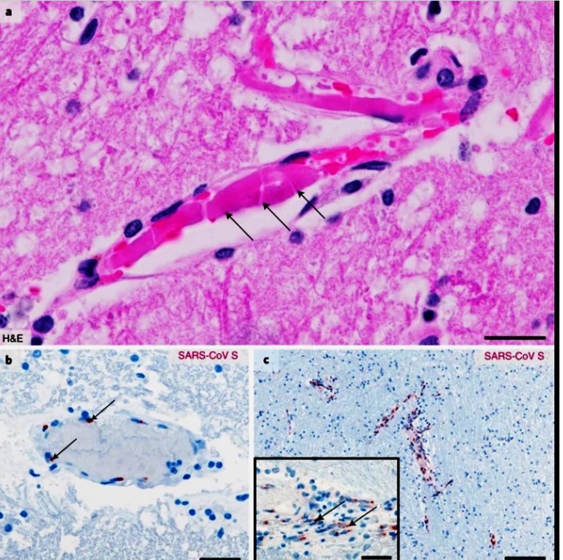
a, Hematoxylin and eosin (H&E)-stained FFPE section of the thalamus obtained from a deceased individual with COVID-19 (individual P26). Several small vessels exhibit fresh thrombi (pink, indicated by arrows) resulting in a large infarct of surrounding CNS tissue characterized by a substantial reduction of detectable neuronal and glial nuclei, edema and vacuolation. b,c, SARS-CoV S protein observed in the endothelial cells of small CNS vessels. Tissue with no obvious ischemic damage exhibits only sparse staining intensity in endothelial cells (b, medulla oblongata, n = 3 of 6; red, indicated by arrows, individual P3) when compared to endothelial cells within acute infarct areas (c, pons, n = 3 of 4; red, indicated by arrows, individual P4; inset depicts a magnified vessel from a different region of the same specimen exhibiting SARS-CoV S protein deposits within endothelial cells). Scale bars: 30 µm (a), 50 µm (b), 200 µm (c) and 40 µm (inset in c).
https://www.nature.com/articles/s41593-020-00758-5