

COVID-19 Clinical Cases:Pulmonary Embolism
Case presented at the NEJM360
Hugo Juan Gallinari. M.D. Ph.D. Cardiology and Family Practice, HCC Panaderas, Madrid
A 36-year-old man, without history of any chronic disease or other clinical infection and without antecedent of substance-use or mental disorder, was admitted to the hospital because of fever, dyspnoea, chest pain and cough.
The patient had been in her usual state of health until approximately 10 days before admission, when he reportedly had a high fever that no resolved after 4 days. Six days before admission and daily thereafter, his temperature rose as high as 40°C, with associated chills, rigors, and pain in the chest, low back, and majority of joints. On the evening before admission, the temperature was reportedly 38.9°C after the administration of ibuprofen 600 mg – acetominophen 1000 mg.. The next afternoon, he came to the emergency department of his reference hospital for evaluation.
On examination, the patient was alert and fully oriented. The temperature was 37.8°C, the blood pressure 125/72 mm Hg, the pulse 107 beats per minute, the respiratory rate 23 breaths per minute, and the oxygen saturation 92% while he was breathing ambient air. The heart sounds were normal, without murmurs. No Inguinal or others lymphadenopathy was noted, without a clinical evidence of abscesses or infection. Joints not were warm. The spine was tender between the T12 and the L3 vertebrae, without paraspinal discomfort. The remainder of the examination was normal. An electrocardiogram showed sinus tachycardia at a rate of 107 beats per minute and was otherwise normal.
Analytical at admission show alterations such as lymphopenia, low elevation of transaminases and lactate dehydrogenase. Elevation of D Dimer Over 800 mcg/L, and ferritin of 1300 mcg/L, without impairment of King. Elevation of CK was detected with no elevated troponopin. Echocardiography was normal.
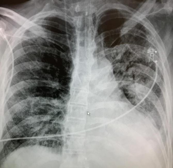
Postero-anterior chest radiographs showed low inspiration. The ground glass patterned areas, which affectation of both lungs, in particular the lower lobes, and especially the posterior segments.
Computed tomography (CT) of the chest was performed and suggest more diffuse pattern.
Positive PCR test was performed at admission, and was positive for COVID-19 infection.
Was treated at admission with Hydroxychloroquin 400 mg bid first day and 200 mg bid after first day, and azithromycin 500 mg bid, oxygen and aerosol therapy.
At 3 days, patient show an impairment of king and liver function, with alteration of coagulation, and deteriorates of lung images (lesions progress until they become more diffuse) and oxygenation. Was incubated in prone positioning.
Patient show at 4 days of admission, Suddenly respiratory failure not fully explained by cardiac failure or fluid overload, with low oxygenation. Urgent TC show bilateral pulmonary thrombo-embolism, and no evidence of deep vein thrombosis but with reduced cardiac function. Was started treatment with heparin, but died at night of the 4 day. And this is the second younger patient with this evolution in the last week.